A) suture.
B) syndesmosis.
C) gomphosis.
D) synostosis.
E) symphysis.
G) A) and B)
Correct Answer

verified
Correct Answer
verified
Multiple Choice
A synchondrosis
A) is a type of gomphosis.
B) is freely movable.
C) may be temporary.
D) is found in the arm.
E) is not found in a growing long bone.
G) A) and B)
Correct Answer

verified
Correct Answer
verified
Multiple Choice
Delores is seeing a neurologist for severe inflammation in a specific type of joint.Which of these joints would be most likely to cause problems with the spinal cord?
A) cubital joint
B) glenohumeral joint
C) atlantoaxial joint
D) sternoclavicular joint
E) talocrural joint
G) D) and E)
Correct Answer

verified
Correct Answer
verified
Multiple Choice
The medial meniscus is in the
A) neck.
B) shoulder.
C) hip.
D) knee.
E) elbow.
G) A) and C)
Correct Answer

verified
Correct Answer
verified
Multiple Choice
Synovial fluid
A) lacks cells.
B) is found between all bony junctions.
C) increases friction between bones.
D) is produced by articular cartilage.
E) decreases friction between bones.
G) B) and C)
Correct Answer

verified
Correct Answer
verified
Multiple Choice
The inability to produce the fluid that keeps most joints moist indicates a disorder of the
A) cruciate ligament.
B) synovial membrane.
C) articular cartilage.
D) bursae.
E) mucus membrane.
G) A) and E)
Correct Answer

verified
Correct Answer
verified
Multiple Choice
Which of the following movements is possible at the hip or coxal joint?
A) rotation
B) flexion
C) adduction
D) circumduction
E) All of these are possible.
G) A) and C)
Correct Answer

verified
Correct Answer
verified
Multiple Choice
Yolanda,a yoga instructor,tells her class to stretch the muscles of the side of the trunk by instructing them to perform ___.
A) opposition.
B) adduction.
C) lateral flexion.
D) extension.
E) elevation.
G) A) and E)
Correct Answer

verified
C
Correct Answer
verified
Multiple Choice
The type of movement between carpal bones is described as
A) pivot.
B) adduction.
C) extension.
D) flexion.
E) gliding.
G) A) and B)
Correct Answer

verified
Correct Answer
verified
Multiple Choice
Arthritis is
A) a bacterial infection transmitted by ticks.
B) an inflammation of any joint.
C) a metabolic disorder caused by increased uric acid in blood.
D) a condition that may involve an autoimmune disease.
E) the most common type of arthritis.
G) All of the above
Correct Answer

verified
Correct Answer
verified
Multiple Choice
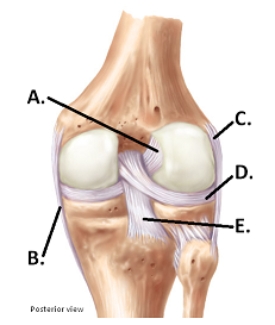
 -The figure illustrates a posterior view of the right knee joint.What does "A" represent?
A) medial (tibial) collateral ligament (MCL)
B) posterior cruciate ligament (PCL)
C) anterior cruciate ligament (ACL)
D) lateral (fibular) collateral ligament (LCL)
E) lateral meniscus
G) D) and E)
Correct Answer

verified
Correct Answer
verified
Multiple Choice
A movement through 360 degrees that combines flexion,extension,abduction,and adduction is called
A) circumduction.
B) rotation.
C) hyperextension.
D) supination.
E) pronation.
G) A) and C)
Correct Answer

verified
A
Correct Answer
verified
Multiple Choice
Gout is
A) a bacterial infection transmitted by ticks.
B) an inflammation of any joint.
C) a metabolic disorder caused by increased uric acid in blood.
D) a condition that may involve an autoimmune disease.
E) the most common type of arthritis.
G) C) and D)
Correct Answer

verified
Correct Answer
verified
Multiple Choice
 -The figure illustrates a posterior view of the right knee joint.What does "B" represent?
A) medial (tibial) collateral ligament (MCL)
B) posterior cruciate ligament (PCL)
C) anterior cruciate ligament (ACL)
D) lateral (fibular) collateral ligament (LCL)
E) lateral meniscus
G) B) and D)
Correct Answer

verified
Correct Answer
verified
Multiple Choice
 -The figure illustrates a posterior view of the right knee joint.What does "D" represent?
A) medial (tibial) collateral ligament (MCL)
B) posterior cruciate ligament (PCL)
C) anterior cruciate ligament (ACL)
D) lateral (fibular) collateral ligament (LCL)
E) lateral meniscus
G) C) and D)
Correct Answer

verified
Correct Answer
verified
Multiple Choice
Rotating the forearm so that the palm faces posteriorly is called
A) circumduction.
B) rotation.
C) hyperextension.
D) supination.
E) pronation.
G) A) and D)
Correct Answer

verified
Correct Answer
verified
Multiple Choice
 -The figure illustrates a posterior view of the right knee joint.What does "E" represent?
A) medial (tibial) collateral ligament (MCL)
B) posterior cruciate ligament (PCL)
C) anterior cruciate ligament (ACL)
D) lateral (fibular) collateral ligament (LCL)
E) lateral meniscus
G) None of the above
Correct Answer

verified
Correct Answer
verified
Multiple Choice
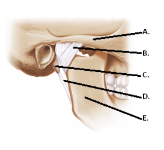
 -The figure illustrates structures in the right temporomandibular joint (lateral view) .What does "C" represent?
A) lateral ligament
B) mandible
C) zygomatic arch
D) styloid process
E) stylomandibular ligament
G) A) and C)
Correct Answer

verified
Correct Answer
verified
Multiple Choice
Turning the ankle so that the plantar surface faces laterally is
A) eversion.
B) inversion.
C) supination.
D) retraction.
F) A) and D)
Correct Answer

verified
Correct Answer
verified
Multiple Choice
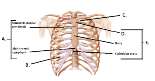
 -The figure illustrates the joints and bones of the rib cage.What does "A" represent?
A) costochondral joint
B) sternum
C) manubrium
D) sternal symphyses
E) sternocostal synchrondrosis
G) A) and C)
Correct Answer

verified
D
Correct Answer
verified
Showing 1 - 20 of 119
Related Exams