A) anterior fontanel
B) posterior fontanel
C) parietal bone
D) sagittal suture
E) occipital bone
G) A) and C)
Correct Answer

verified
Correct Answer
verified
Multiple Choice
The anterior cruciate ligament prevents _____ displacement of the tibia.
A) anterior
B) posterior
C) lateral
D) medial
E) radial
G) C) and D)
Correct Answer

verified
Correct Answer
verified
Multiple Choice
An example of a symphysis is the
A) elbow joint.
B) temporomandibular joint.
C) costovertebral joint.
D) intervertebral joint.
E) sacroiliac joint.
G) D) and E)
Correct Answer

verified
Correct Answer
verified
Multiple Choice
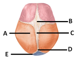
 -The figure illustrates bones,fontanels,and sutures on the fetal skull.What does "E" represent?
A) anterior fontanel
B) posterior fontanel
C) parietal bone
D) sagittal suture
E) occipital bone
G) D) and E)
Correct Answer

verified
Correct Answer
verified
Multiple Choice
Abnormal forced extension beyond normal range of motion is called
A) circumduction.
B) rotation.
C) hyperextension.
D) supination.
E) pronation.
G) B) and C)
Correct Answer

verified
Correct Answer
verified
Multiple Choice
Rheumatoid arthritis is
A) a bacterial infection transmitted by ticks.
B) an inflammation of any joint.
C) a metabolic disorder caused by increased uric acid in blood.
D) a condition that may involve an autoimmune disease.
E) the most common type of arthritis.
G) C) and D)
Correct Answer

verified
Correct Answer
verified
Multiple Choice
A sharp object penetrated a synovial joint.From the following list of structures,select the order in which they were penetrated. (1) tendon or muscle (2) ligament (3) fibrous capsule (4) skin (5) synovial membrane
A) 4,1,2,5,3
B) 4,5,1,2,3
C) 4,3,2,5,1
D) 4,1,2,3,5
E) 4,2,1,5,3
G) C) and D)
Correct Answer

verified
Correct Answer
verified
Multiple Choice
A pivot joint
A) is a modified ball and socket joint.
B) restricts movement to rotation.
C) is a biaxial joint.
D) allows gliding movement.
E) is between the atlas and the occipital bone.
G) C) and E)
Correct Answer

verified
Correct Answer
verified
Multiple Choice
Which of the following statements concerning the ankle joint is true?
A) The calcaneus articulates with the tibia to form this joint.
B) Most common injuries to this joint occur because of a forceful inversion of the foot.
C) A capsule of hyaline cartilage surrounds the joint.
D) The lateral collateral ligament helps to stabilize this joint.
E) It is a pivot joint.
G) A) and B)
Correct Answer

verified
Correct Answer
verified
Multiple Choice
 -The figure illustrates bones,fontanels,and sutures on the fetal skull.What does "B" represent?
A) anterior fontanel
B) posterior fontanel
C) parietal bone
D) sagittal suture
E) occipital bone
G) A) and C)
Correct Answer

verified
Correct Answer
verified
Multiple Choice
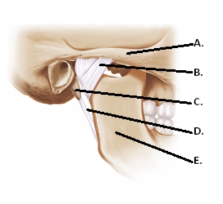
 -The figure illustrates structures in the right temporomandibular joint (lateral view) .What does "E" represent?
A) lateral ligament
B) mandible
C) zygomatic arch
D) styloid process
E) stylomandibular ligament
G) B) and D)
Correct Answer

verified
Correct Answer
verified
Multiple Choice
The opposite of supination is
A) inversion.
B) protraction.
C) elevation.
D) pronation.
E) flexion.
G) B) and D)
Correct Answer

verified
Correct Answer
verified
Multiple Choice
Which of the following movements does not occur at the knee joint?
A) flexion
B) rotation
C) abduction
D) extension
E) All occur at the knee.
G) B) and E)
Correct Answer

verified
Correct Answer
verified
Multiple Choice
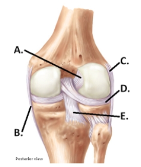
 -The figure illustrates a posterior view of the right knee joint.What does "C" represent?
A) medial (tibial) collateral ligament (MCL)
B) posterior cruciate ligament (PCL)
C) anterior cruciate ligament (ACL)
D) lateral (fibular) collateral ligament (LCL)
E) lateral meniscus
G) B) and E)
Correct Answer

verified
Correct Answer
verified
Multiple Choice
In a syndesmosis
A) there is an osseous union between the bones of the joint.
B) the bones are held together by ligaments called interosseous membranes.
C) it is not unusual to find discs of cartilage.
D) no movement occurs.
E) there is a great range of motion.
G) B) and D)
Correct Answer

verified
Correct Answer
verified
Multiple Choice
A biaxial joint has movement
A) around one axis.
B) around two axes at right angles to one another.
C) about several axes.
D) as long as there is articular cartilage present.
E) that always rotates.
G) B) and D)
Correct Answer

verified
Correct Answer
verified
Multiple Choice
Returning the thumb to the anatomical position after touching the little finger is
A) reposition.
B) opposition.
C) medial excursion.
D) supination.
F) None of the above
Correct Answer

verified
Correct Answer
verified
Multiple Choice
 -The figure illustrates structures in the right temporomandibular joint (lateral view) .What does "A" represent?
A) lateral ligament
B) mandible
C) zygomatic arch
D) styloid process
E) stylomandibular ligament
G) A) and C)
Correct Answer

verified
Correct Answer
verified
Multiple Choice
What is the most commonly dislocated joint in the body?
A) glenohumeral joint
B) temporomandibular joint
C) humeroulnar joint
D) coxal joint
E) knee joint
G) C) and D)
Correct Answer

verified
Correct Answer
verified
Multiple Choice
Bowing the head is an example of
A) rotation.
B) pronation.
C) flexion.
D) lateral excursion.
E) hyperextension.
G) A) and E)
Correct Answer

verified
Correct Answer
verified
Showing 21 - 40 of 119
Related Exams